Home » Without Label » Upper Thigh Anatomy : Single-leg Split Squats on a Bench | FitnessRX for Women ... : These are the gluteus maximus, gluteus medius, gluteus minimus, and tensor fasciae latae.
Upper Thigh Anatomy : Single-leg Split Squats on a Bench | FitnessRX for Women ... : These are the gluteus maximus, gluteus medius, gluteus minimus, and tensor fasciae latae.
Upper Thigh Anatomy : Single-leg Split Squats on a Bench | FitnessRX for Women ... : These are the gluteus maximus, gluteus medius, gluteus minimus, and tensor fasciae latae.. Finally, the hamstring muscles that run down the back of the thigh start on the bottom of the pelvis. Muscles of the lower limb; Anatomically, it is part of the lower limb. 2, vastus medialis & intermedius muscles. The femur is found in the thigh.
Greater trochanteric pain syndrome, also called trochanteric bursitis or gtps, is an inflammation of the bursa of the greater trochanter. Small and deep muscles which mainly externally rotate the thigh at the hip joint and stabilize the pelvis. Each type of muscle tissue in the human body has a unique structure and a specific role. Sartorius, and the four quadriceps muscles; This section of the website will explain large and minute details of arterial anatomy of upper legs (thigh arteries)
Muscles of the Thigh and Gluteal Region - Part 1 - Anatomy ... from i.ytimg.com These are the gluteus maximus, gluteus medius, gluteus minimus, and tensor fasciae latae. Human muscles · july 20, 2016. 2, vastus medialis & intermedius muscles. There are five muscles in the anterior thigh compartment: Ebraheim's educational animated video describes muscle anatomy of the thigh. The femur is found in the thigh. The hamstring muscles, also known as the rear thighs, make up the backside of the upper leg anatomy. Muscles of the lower limb;
It is the largest bone in the body and is the only bone in the upper leg.
Sartorius, and the four quadriceps muscles; The rectus femoris is located in the center of the thigh, while the vastus medialis is in the middle of the said body part. Muscles are named according to their shape, location, or a combination. Knee assessment and hip mechanics online course: Muscle anatomy of upper thigh. 1 article features images from this case. In clinical anatomy the thigh muscles are divided into three groups: 5.0 based on 12 ratings, 7 reviews. It is also referred to as a ball and socket joint and is surrounded by muscles, ligaments, and tendons. Thigh the thigh bears much of the load of the body's weight when a person is upright. The muscle is one of the four quadriceps muscles and is the largest muscle of that group. The hamstring muscles, also known as the rear thighs, make up the backside of the upper leg anatomy. Like the adductors, the abductors are also responsible for stabilizing your knees during athletic and everyday movement.
Anatomically, it is part of the lower limb. The muscles in the upper leg power many of our movements. •medial thigh muscles•adductor longus muscle•adductor magnus muscle•adductor. This mri brain cross sectional anatomy tool is absolutely free to use. Any injury or disease of the hip will.
11 factors that differentiate sciatica from hamstring or ... from www.sportsinjurybulletin.com It also is active in maintaining thigh and kneecap position while walking and. Like the adductors, the abductors are also responsible for stabilizing your knees during athletic and everyday movement. (there are four types of bone: Atlas of body sections, ct and mri images, fourth edition. Bursae are the small cushions between tendons, bones, and. Pretty self explanatory from the title i think. On the anterior side, the most prominent of the muscles are the sartorius muscle and the four muscles that make up quadriceps muscle group (the quads.) It's the area that runs from the hip to the knee in each leg.
Anatomically, it is part of the lower limb.
Medial muscles adduct and rotate your thigh, and posterior flex your leg and extend your thigh. These are the gluteus maximus, gluteus medius, gluteus minimus, and tensor fasciae latae. Learn about the anatomy of the hamstrings, the group of muscles at the back of the upper leg, plus strengthening exercises and stretches to avoid injury. Anterior muscles extend your legs and flex your thighs. This section of the website will explain large and minute details of arterial anatomy of upper legs (thigh arteries) Anatomically, it is part of the lower limb. The muscles in the upper leg power many of our movements. Each type of muscle tissue in the human body has a unique structure and a specific role. Muscles are named according to their shape, location, or a combination. Case contributed by dr roberto schubert. The femur is found in the thigh. Long bones, short bones, flat bones, and irregular bones.) long bones are longer than they are wide, with spongy bones at both ends and a cavity filled with bone. (there are four types of bone:
The four muscles all extend the lower leg. Atlas of body sections, ct and mri images, fourth edition. Abductors are located on the upper portion of the outside of your thighs and hips, anchoring above on the pelvis, and below at various points on your outside thigh. The rectus femoris is located in the center of the thigh, while the vastus medialis is in the middle of the said body part. Tools:wacom intuos3 tabletadobe photoshopcamtasia studiomusic by mefor free art resources, like photoshop br.
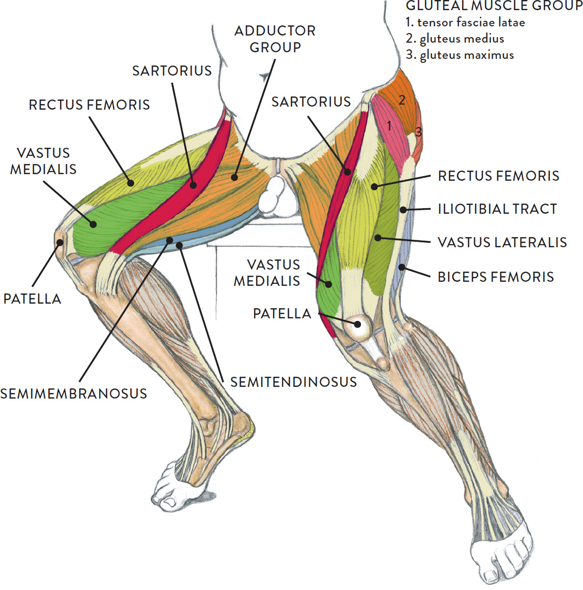
Graphite pencil, watercolor wash, and cream and white ... from schoolbag.info In clinical anatomy the thigh muscles are divided into three groups: 1 article features images from this case. Like the forearm, the upper leg, or thigh, has a dense arrangement of many muscles. Rectus femoris, vastus medialis, vastus lateralis and vastus intermedius. Abductors are located on the upper portion of the outside of your thighs and hips, anchoring above on the pelvis, and below at various points on your outside thigh. The vastus lateralis is a muscle located on the lateral, or outside, part of your thigh. The rectus femoris is located in the center of the thigh, while the vastus medialis is in the middle of the said body part. Ebraheim's educational animated video describes muscle anatomy of the thigh.
2, vastus medialis & intermedius muscles.
The vastus lateralis is a muscle located on the lateral, or outside, part of your thigh. Like the adductors, the abductors are also responsible for stabilizing your knees during athletic and everyday movement. One further muscle of the anterior knee is the small articularis genus muscle, it is occasionally is blended with vastus intermedius. Any injury or disease of the hip will. The posterior upper leg muscles provide your knees with mobility (extension, flexion and rotation) and strength. Along the upper portion of the thigh, just lateral to the gracilis, the adductor longus muscle is ranked as the most anterior of this group of thigh muscles upper thigh anatomy. In clinical anatomy the thigh muscles are divided into three groups: It also is active in maintaining thigh and kneecap position while walking and. Diagnosis not applicable diagnosis not applicable. Ebraheim's educational animated video describes muscle anatomy of the thigh. Small and deep muscles which mainly externally rotate the thigh at the hip joint and stabilize the pelvis. •medial thigh muscles•adductor longus muscle•adductor magnus muscle•adductor. By spicer mcleroy in tutorials.